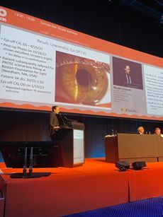
Dr. Mabel Cheng and Dr. Nicole Lemanski introduce Epithelial Mapping and Widefield Digital Angiography
In a first for patients in the Capital Region, Dr. Mabel Cheng and Dr. Nicole Lemanski are proud to announce the introduction of high resolution anterior segment OCT with epithelial mapping and Widefield Retinal Digital Angiography.
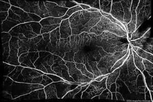
“These two upgrades, released by Zeiss in November 2019, greatly expand the diagnostic capacity afforded to our patients,” states Brian Lemanski, practice administrator. “Digital OCT Angiography allows us to conduct vascular studies of the back of the eye that usually required a dye solution to be injected into a patient. Now a fair majority can be performed using just light, making it easier for hassle free routine imaging. The widefield aspect is equally important, as it affords examination of a larger portion of the retina, making it more likely for disease processes to be detected. Diabetics in particular, who are recommended to have a dilated eye exam at least once a year, will greatly benefit from this technology to look for more subtle diabetic eye disease.”

High Resolution Epithelial Mapping (image courtesy of Zeiss).
“Meanwhile, the epithelium, the outer-most layer of the clear part of the eye, is poorly visualized with traditional imaging techniques. High resolution epithelial mapping greatly enhances our understanding of certain corneal pathologies, and affords more tailored treatments for conditions such as recurrent erosion and enhanced detection of sub-clinical keratoconus.”
Have a question on any of our technologies? Feel free to contact us at 518-782-7777 for more details.
–The office staff of Dr. Mabel Cheng and Dr. Nicole Lemanski